Gliolan

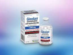
Gliolan is an innovative pharmaceutical agent utilized during neurosurgical procedures to enhance the visibility and excision of malignant tissues. Its active ingredient is 5-aminolevulinic acid hydrochloride (5-ALA HCl). This remarkable compound is particularly advantageous in the surgical management of high-grade gliomas, including glioblastoma, a notably aggressive form of brain tumor. By functioning as a fluorescent imaging enhancer, Gleolan enables surgeons to differentiate between neoplastic and healthy brain tissues with remarkable precision.
How Does Gleolan Function?
Administered orally prior to surgery, Gleolan is absorbed into the bloodstream and subsequently transformed into protoporphyrin IX (PpIX) within the cellular environment. Tumor cells exhibit a higher affinity for 5-ALA, converting it into PpIX in significantly greater quantities than their normal counterparts. During the surgical procedure, when exposed to a specific blue light, the PpIX within the tumor cells emits a striking pink or red fluorescence, while the surrounding healthy brain tissue remains unaffected. This luminous distinction greatly aids surgeons in excising the tumor while preserving vital healthy tissue.
Applications of Gleolan
Gleolan is primarily employed in neurosurgical interventions for patients diagnosed with:
High-grade Gliomas: This category encompasses glioblastoma, characterized by its rapid proliferation and challenging treatment landscape.
Other Malignant Brain Tumors: Gleolan proves beneficial in visualizing various malignant neoplasms, thereby enhancing surgical efficacy.
The overarching objective of utilizing Gleolan is to maximize tumor resection while safeguarding healthy brain structures. Achieving complete tumor removal is often associated with improved prognoses, including extended survival rates and diminished recurrence risks.
How is Gleolan Administered?
Gleolan is provided as a palatable solution approximately three hours prior to the surgical procedure. The dosage is tailored to the patient’s weight, typically around 20 mg of 5-ALA per kilogram. Following ingestion, the medication requires a brief period for absorption and metabolic conversion to ensure optimal effectiveness during the surgical intervention.
1.Advantages of Gleolan
Enhanced Tumor Visualization: Gleolan significantly improves the surgeon’s ability to visualize tumors, allowing for a clear distinction between malignant growths and healthy brain tissue.
Increased Tumor Removal: The use of Gleolan facilitates a more thorough excision of tumors, which is associated with improved patient prognoses.
Better Preservation of Brain Function: By precisely locating the tumor, surgeons can safeguard vital brain areas, thereby minimizing the likelihood of neurological complications.
Potential Side Effects
Though generally regarded as safe, Gleolan may lead to certain side effects:
Photosensitivity: Patients may experience heightened sensitivity to light for up to 48 hours post-administration. It is advisable to steer clear of direct sunlight and intense indoor lighting during this time.
Nausea and Vomiting: Some individuals may encounter mild to moderate nausea or vomiting following the medication.
Headache: Mild headaches may manifest as a side effect.
Drop in Blood Pressure: A temporary decrease in blood pressure may occur in some patients during or after the surgical procedure.
Safety Precautions
Avoid Bright Light: To mitigate skin and eye irritation, patients should refrain from exposure to sunlight. Bright artificial lighting for a minimum of 24 hours after taking Gleolan.
Medical History: It is crucial for patients to disclose any pre-existing medical conditions to their physician, particularly those related to liver health or any medications that may influence liver function.
Allergic Reactions: Although infrequent, allergic reactions to Gleolan can happen. Patients should promptly inform their healthcare provider of any unusual symptoms.
Conclusion
Gleolan serves as an indispensable asset in the realm of brain surgery, especially for individuals diagnosed with high-grade gliomas. By illuminating tumor cells under specific lighting conditions, it empowers surgeons to excise tumors with greater efficacy and safety. This not only enhances patient outcomes but also diminishes the risk of tumor recurrence. Aids in the preservation of brain function. Patients are encouraged to adhere to all guidelines meticulously. Engage in open dialogue with their healthcare team to optimize the results of their surgical experience.


